Teaching Sets
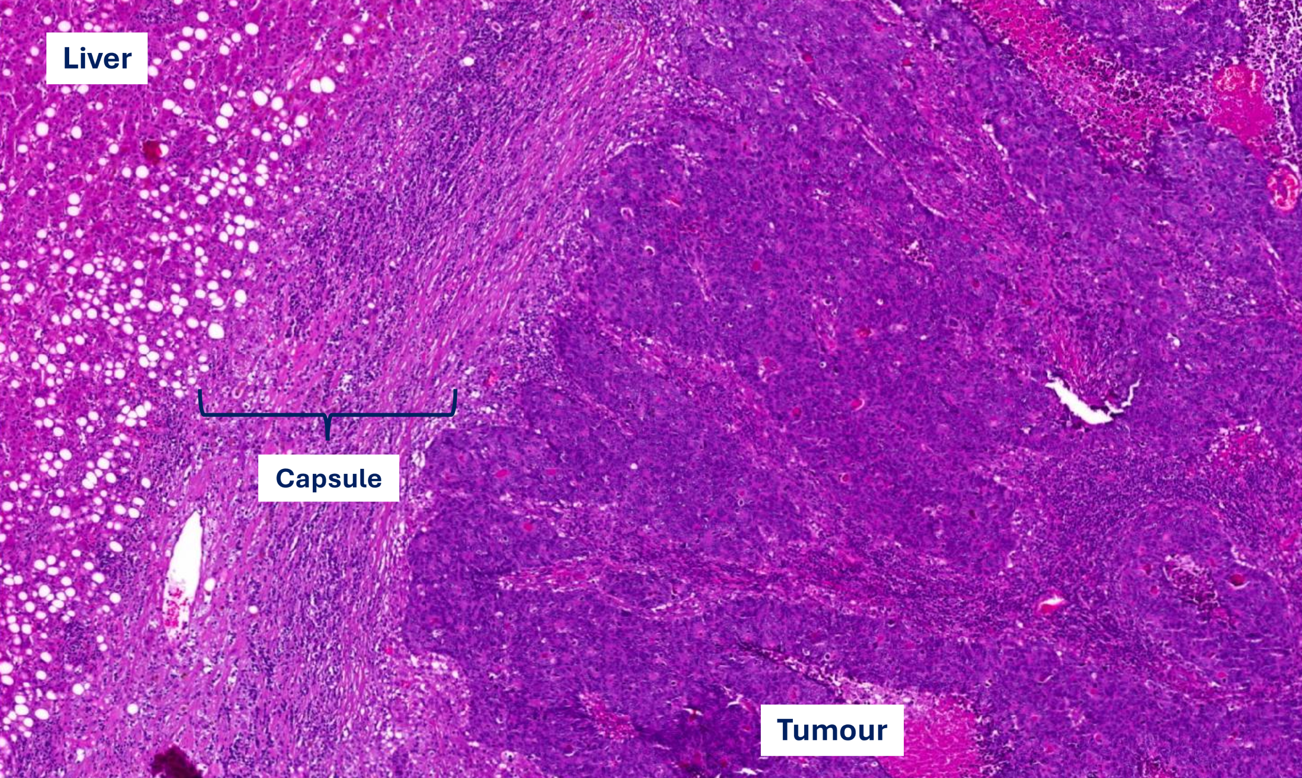
Encapsulated liver metastasis (colorectal cancer)
The tumour (well to moderately differentiated) is separated from the liver by a broad capsule with an inner and outer layer. The outer layer contains a dense immune cell infiltrate extending to the liver. The inner layer consists of compact connective tissue with less immune cells.
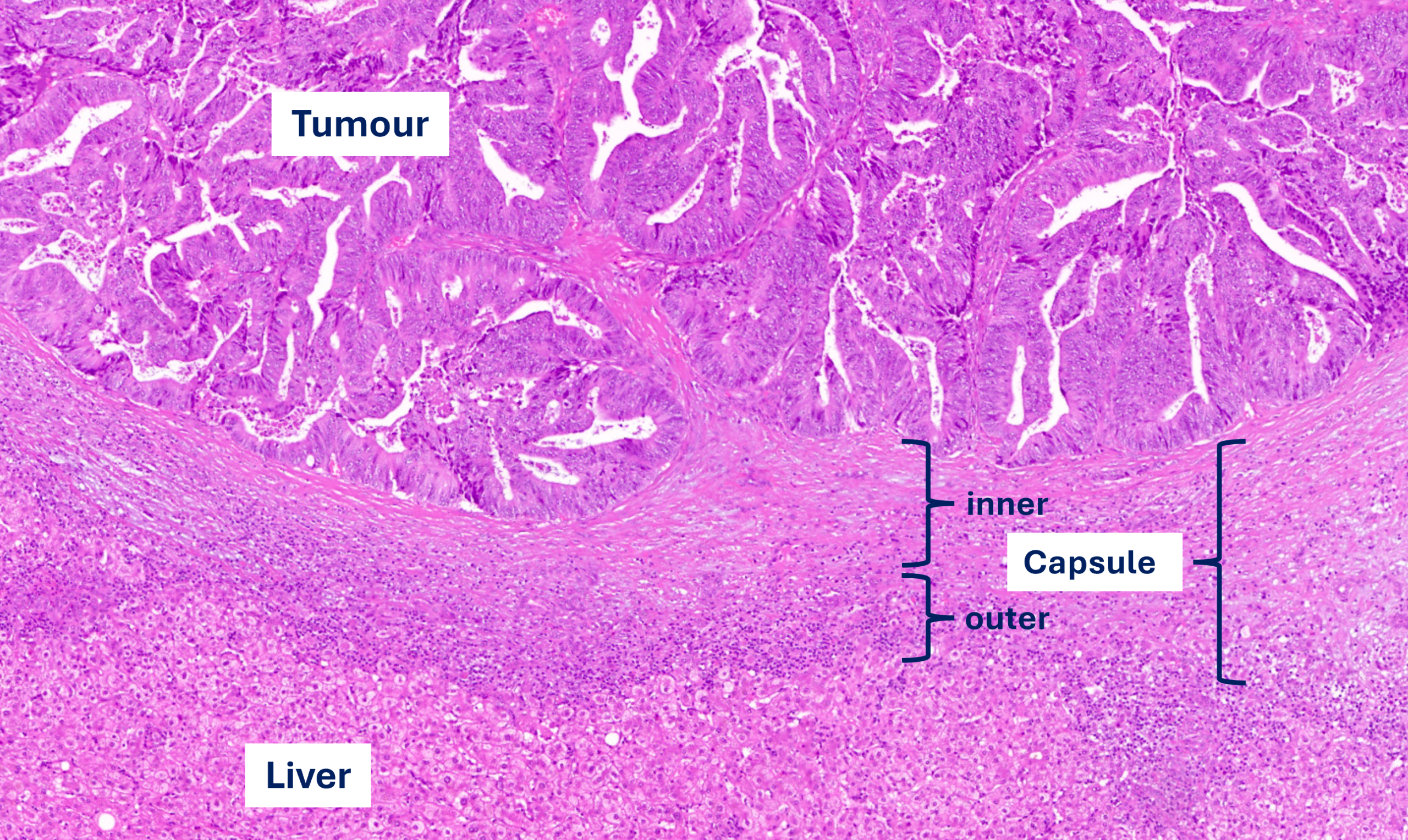
Replacement liver metastasis (colorectal cancer)
The tumour (poorly differentiated) is not separated from the liver. Cancer cells are in contact with hepatocytes (arrows at interface) and hepatocytes are taken up by the tumour (arrows deeper in the tumour), as well as the sinusoidal blood vessels (arrow heads).
There are only few immune cells at the tumour-liver interface.
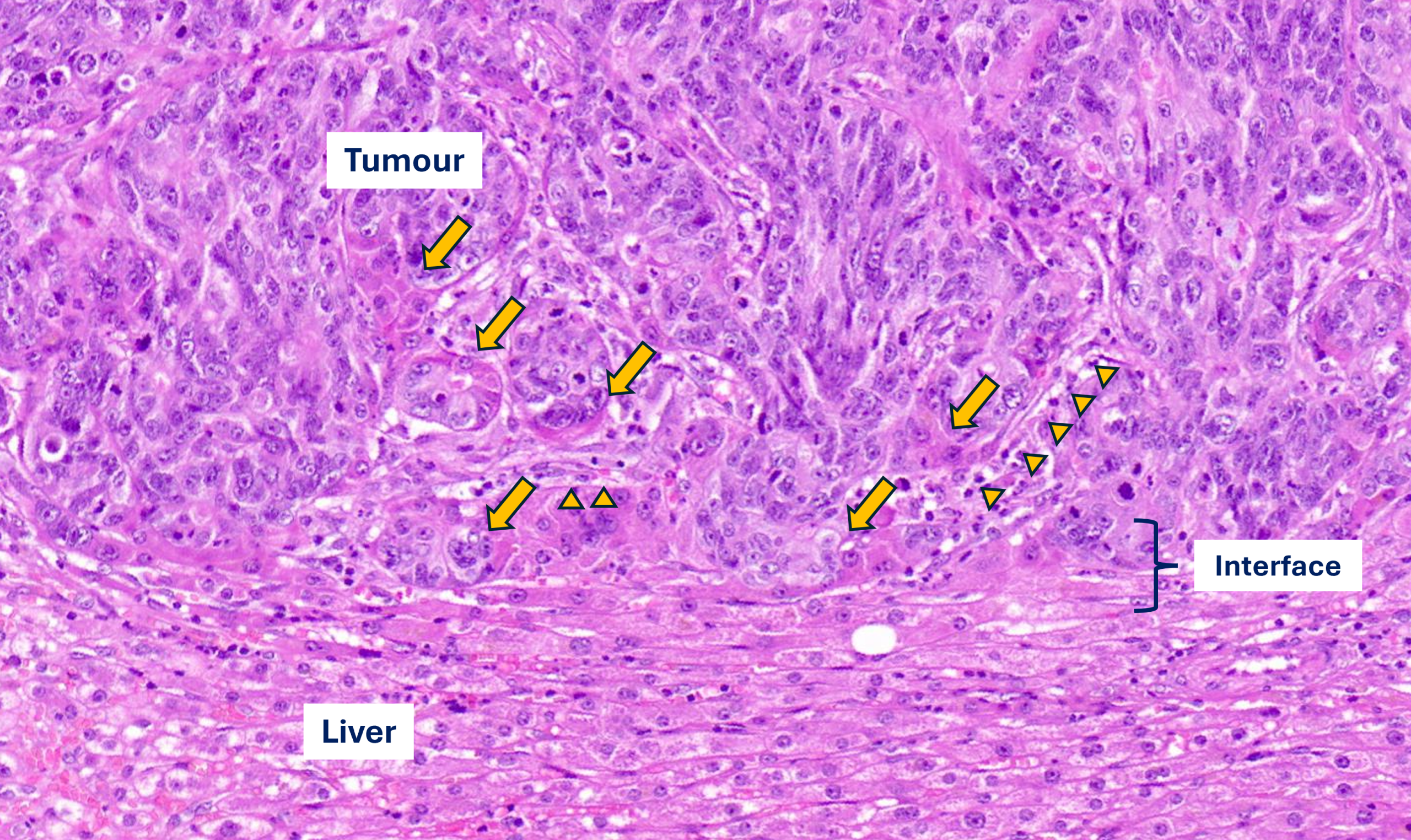
Replacement liver metastasis (colorectal cancer)
The tumour is forming pseudoglandular structures as an expression of differentiation, even at the tumour-liver interface. Yet, cancer cells are in contact with hepatocytes (arrows at interface) and form more solid nests when interacting with the hepatocytes.
There is a moderately dense immune cell infiltrate at the tumour-liver interface.
The connective tissue stroma in between the pseudoglandular structures has the same loose aspect as the stroma in between the liver cell plates (green asterisks), indicating that there is only minimal fibrosis going on during co-option of the sinusoidal blood vessels.
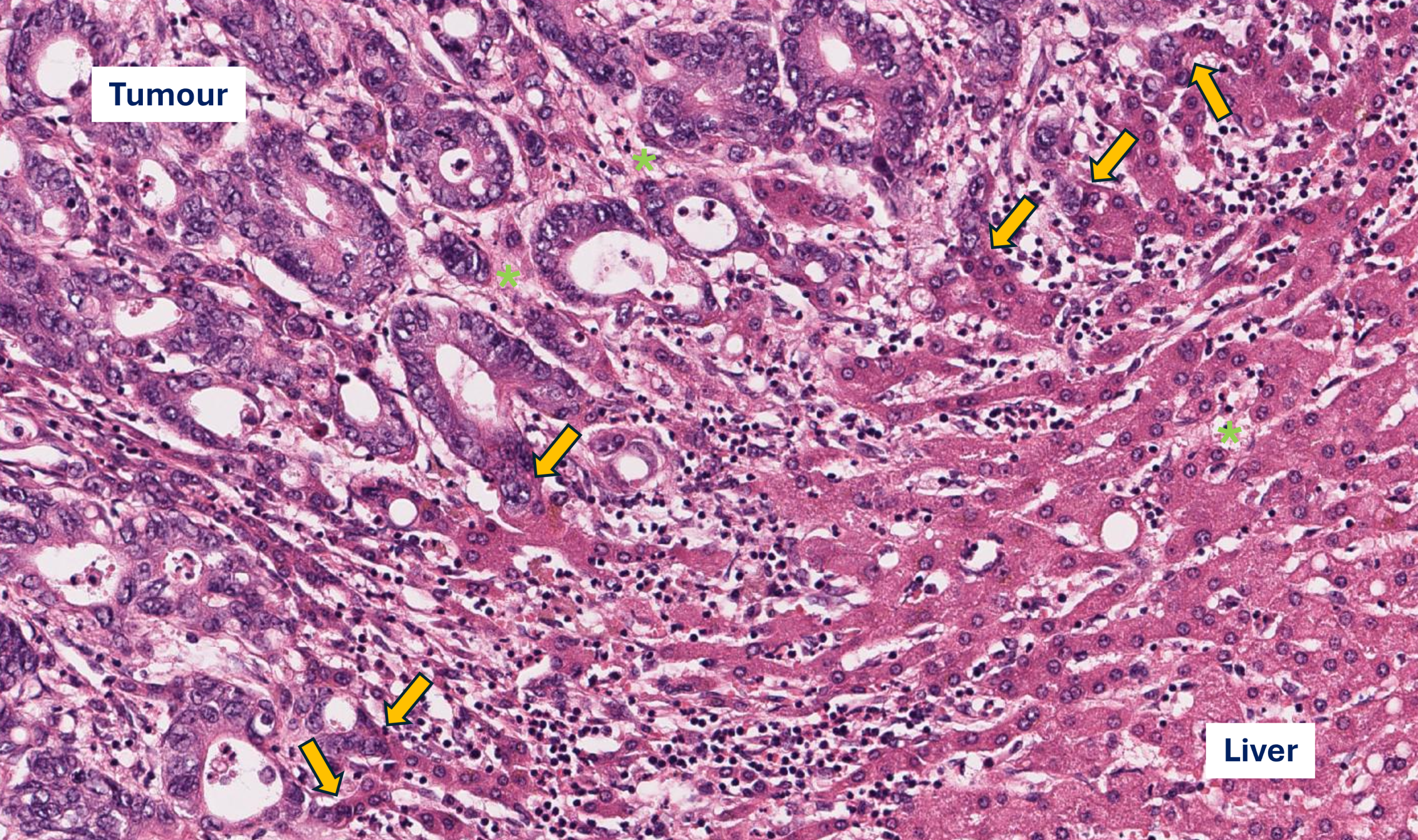
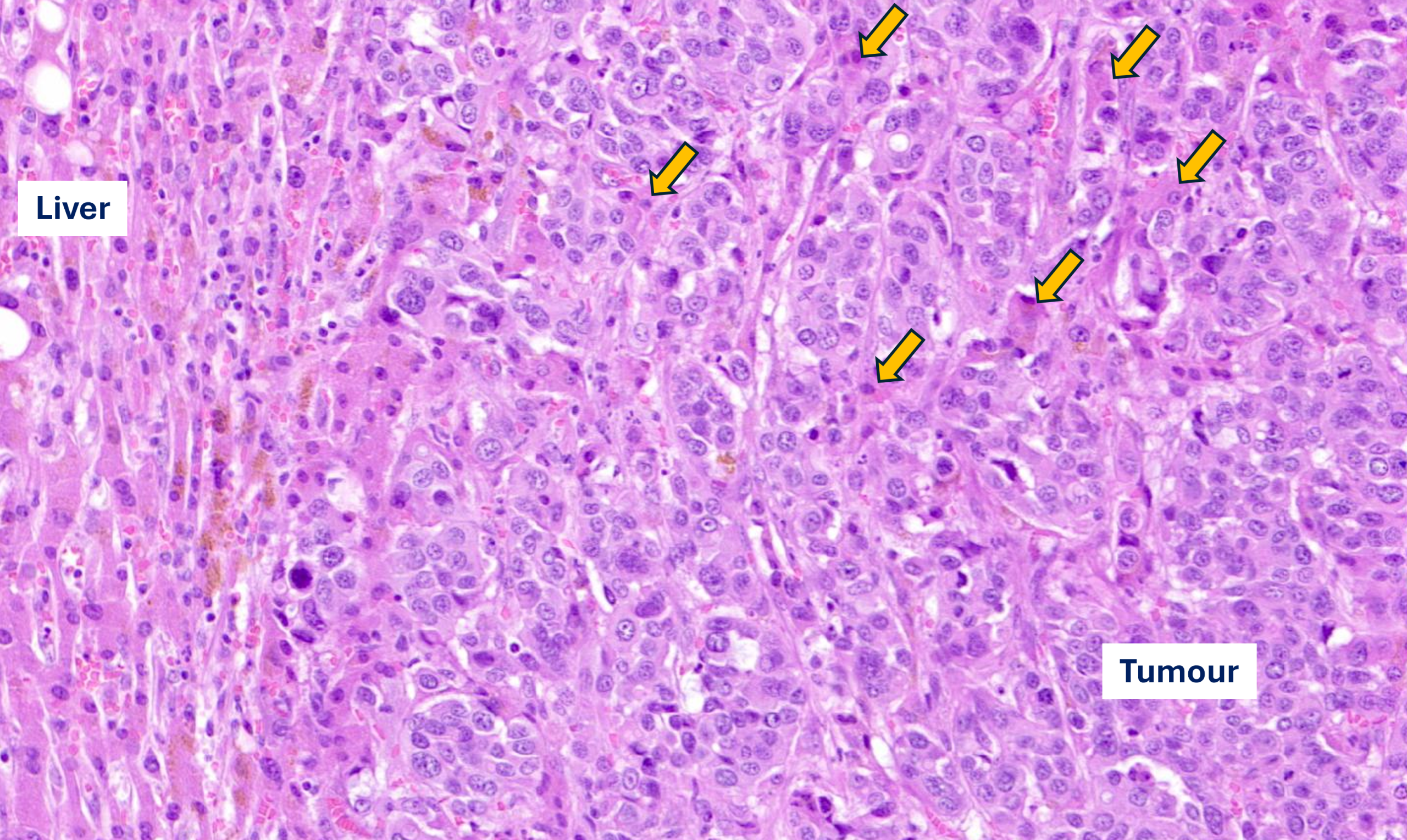
Replacement liver metastasis (colorectal cancer)
Poorly differentiated metastasis with cancer cells arranged in small solid nests, clearly touching hepatocytes at the interface. There is no fibrosis and there are only very few immune cells.
The arrows denote hepatocytes that are taken up by the tumour during the process of vessel co-option.
There are only few immune cells at the tumour-liver interface.
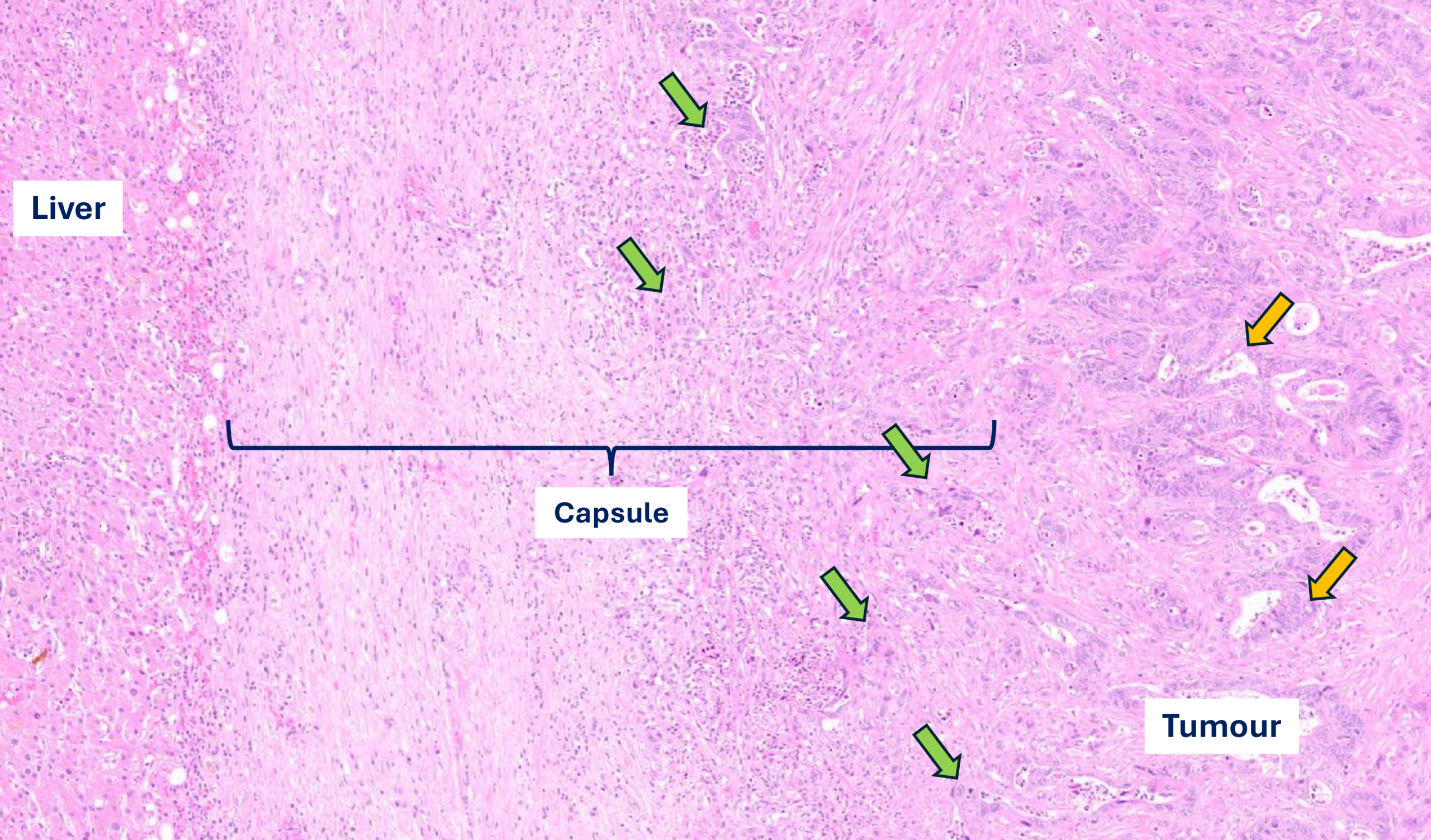
Encapsulated liver metastasis (colorectal cancer)
Moderately differentiated metastasis with cancer cells arranged in small pseudoglandular structures (yellow arrows) with, towards the capsule, smaller solid nests with invasion of the capsule (green arrows).
Broad capsule without clear layering. Also, minimal inflammation at the liver-capsule interface.
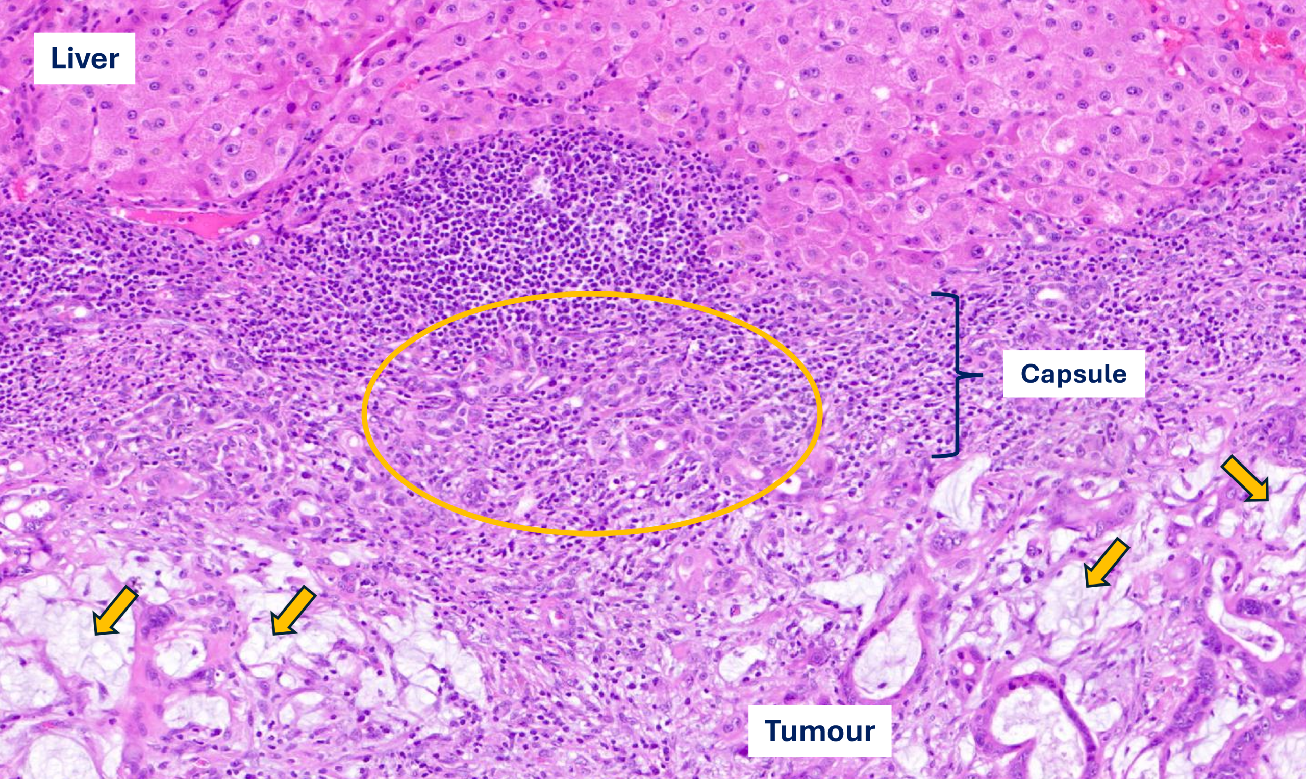
Encapsulated liver metastasis (colorectal cancer – chemotherapy before resection)
Mucinous change, due to systemic treatment, in the metastasis (yellow arrows).
Dense immune cell infiltrate in the fibrotic capsule and extensive ductular proliferation (yellow oval).
Encapsulated liver metastasis (colorectal cancer)
Broad capsule surrounding a poorly differentiated metastasis.
Dense immune cell infiltrate, mainly in the outer half of the capsule.
